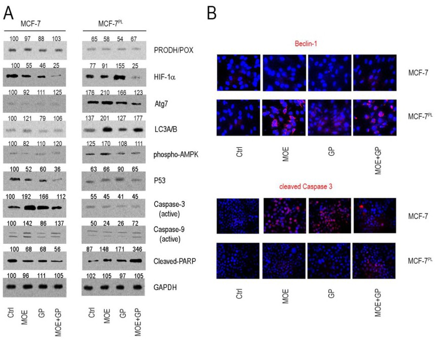
Fig. 5. Western immunoblot (A) for PRODH/POX, autophagy markers (HIF-1α, Atg 7, LC3A/B, phospho-AMPK, beclin-1) and apoptosis markers (p53, cleaved caspase-3 and -9, cleaved PARP) and immunofluorescence analysis (B) for beclin-1 and cleaved Caspase-9 in wild type MCF-7 and MCF-7PL cells untreated (Ctrl) and treated with glycyl-proline (GP), 2-methoxyestradiol (MOE) or MOE + GP in glutamine-free DMEM for 24h. GAPDH was used as a reference protein. The mean values of bands densitometry were added above appropriated bands. All bioimages are included in the Supplementary Material section (see www.cellphysiolbiochem.com).